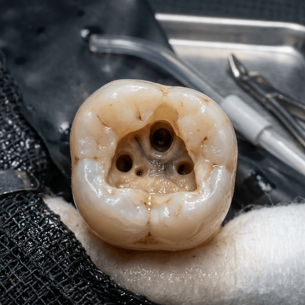
You finish a maxillary first molar root canal. The obturation looks great, the patient is asymptomatic, and everything seems successful. Two years later, the tooth presents with a persistent periapical lesion. What went wrong? The answer may be an untreated MB2 canal. Let’s discuss how and why this happens and important notes that can reduce the likelihood of a missed canal.
A missed canal remains one of the most common causes of endodontic failure, particularly in maxillary first molars. A 2020 cross-sectional study found that canals were missed in approximately 12% of treated teeth, with 62.8% of these missed canals occurring in the mesiobuccal root of maxillary first molars.1
Studies have demonstrated MB2 canal prevalence in maxillary first molars approaching 90% or higher, depending on the detection method used. However, detection remains challenging. One study evaluated MB2 identification using three methods: preoperative CBCT evaluation, direct occlusal access, and coronal grinding. The results demonstrated significant differences between detection methods, showing that MB2 canals may be missed when relying solely on CBCT interpretation, initial access, or both.2
MB2 canals were identified more frequently through direct occlusal access compared with reviewing preoperative CBCT alone. This suggests that routine preoperative CBCT may not be necessary for every maxillary first molar root canal treatment. However, when an MB2 canal cannot be located clinically, CBCT imaging can significantly improve the likelihood of detection and assist in treatment planning.2
The clinical consequences of a missed MB2 canal are significant. In teeth with untreated canals, 82.6% were associated with periapical pathology. Specifically, maxillary first molars with missed MB2 canals demonstrated periapical lesions in 75.2% of cases and were 3.1 times more likely to develop periapical pathology compared with teeth where all canals were identified and obturated.1
Clinical Takeaway:
The MB2 canal should be considered present until proven otherwise. While CBCT is a valuable diagnostic tool, careful access preparation, magnification, and knowledge of root canal anatomy remain essential to reducing the risk of missed anatomy and improving endodontic outcomes.
Resources:
- Baruwa A, Martins J, Meirinhos J …The Influence of Missed Canals on the Prevalence of Periapical Lesions in Endodontically Treated Teeth: A Cross-sectional StudyJournal of Endodontics, 2019; 46, 34-39.e1
- Hiebert B, Abramovitch K, Rice D …Prevalence of Second Mesiobuccal Canals in Maxillary First Molars Detected Using Cone-beam Computed Tomography, Direct Occlusal Access, and Coronal Plane Grinding Journal of Endodontics, 2017; 43, 1711-1715

